[vc_row css_animation=”none”][vc_column css_animation=”fadeInDown” width=”1/4″ offset=”vc_hidden-sm vc_hidden-xs”][vc_wp_custommenu nav_menu=”57″][/vc_column][vc_column css_animation=”fadeInUp” width=”3/4″ offset=”vc_col-lg-offset-0 vc_col-lg-9 vc_col-md-offset-0 vc_col-md-9 vc_col-sm-offset-1″][vc_custom_heading source=”post_title” font_container=”tag:h2|text_align:left|color:%23d71149″ use_theme_fonts=”yes” css=”.vc_custom_1641086513421{margin-bottom: 5px !important;}”][vc_separator color=”custom” align=”align_left” border_width=”2″ el_width=”10″ accent_color=”#eea616″][vc_row_inner][vc_column_inner width=”1/3″][vc_single_image image=”3532″][/vc_column_inner][vc_column_inner width=”2/3″][vc_column_text css=”.vc_custom_1639632768860{margin-bottom: 0px !important;}”]
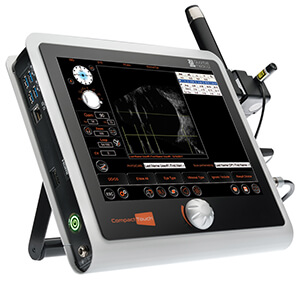
B-scan ultrasonography is commonly used to provide a cross-sectional image of the internal structures of the eye. It is often used to measure tumors, or to detect retinal or choroidal detachment. It is also used when the view of the retina is obscured and can not be examined clearly, such as in cases of vitreous hemorrhage, dense cataract, or in trauma patients. The technique is very quick to perform, non-invasive, and completely safe.
B-scan is commonly used to identify conditions such as,
[/vc_column_text][vntd_icon_list style=”outline” elements=”%5B%7B%22icon_fontawesome%22%3A%22fas%20fa-angle-right%22%2C%22text%22%3A%22Cataract%22%7D%2C%7B%22icon_fontawesome%22%3A%22fas%20fa-angle-right%22%2C%22text%22%3A%22Vitreous%20degeneration%22%7D%2C%7B%22icon_fontawesome%22%3A%22fas%20fa-angle-right%22%2C%22text%22%3A%22Retinal%20detachment%22%7D%2C%7B%22icon_fontawesome%22%3A%22fas%20fa-angle-right%22%2C%22text%22%3A%22Ocular%20trauma%22%7D%2C%7B%22icon_fontawesome%22%3A%22fas%20fa-angle-right%22%2C%22text%22%3A%22Choroidal%20melanoma%22%7D%2C%7B%22icon_fontawesome%22%3A%22fas%20fa-angle-right%22%2C%22text%22%3A%22Retinoblastoma%22%7D%5D” border=”off”][/vc_column_inner][/vc_row_inner][/vc_column][/vc_row]