[vc_row css_animation=”none”][vc_column css_animation=”fadeInDown” width=”1/4″ offset=”vc_hidden-sm vc_hidden-xs”][vc_wp_custommenu nav_menu=”57″][/vc_column][vc_column css_animation=”fadeInUp” width=”3/4″ offset=”vc_col-lg-offset-0 vc_col-lg-9 vc_col-md-offset-0 vc_col-md-9 vc_col-sm-offset-1″][vc_custom_heading source=”post_title” font_container=”tag:h2|text_align:left|color:%23d71149″ use_theme_fonts=”yes” css=”.vc_custom_1641086629240{margin-bottom: 5px !important;}”][vc_separator color=”custom” align=”align_left” border_width=”2″ el_width=”10″ accent_color=”#eea616″][vc_row_inner][vc_column_inner width=”1/3″][vc_single_image image=”3693″ img_size=”medium”][/vc_column_inner][vc_column_inner width=”2/3″][vc_column_text css=”.vc_custom_1639633516299{margin-bottom: 0px !important;}”]
Optical Coherence Tomography is a noninvasive imaging technology used to obtain high resolution cross-sectional images of the retina. The layers within the retina can be differentiated and retinal thickness can be measured to aid in the early detection and diagnosis of retinal diseases and conditions.
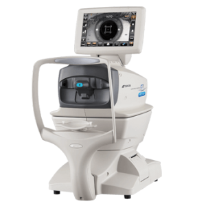
OCT testing has become a standard of care for the assessment and treatment of most retinal conditions. OCT uses rays of light to measure retinal thickness. No radiation or X-rays are used in this test, an OCT scan does not hurt and it is not uncomfortable.
[/vc_column_text][/vc_column_inner][/vc_row_inner][vc_row_inner][vc_column_inner][vc_column_text css=”.vc_custom_1641633670620{margin-bottom: 0px !important;}”]
OCT is useful in diagnosing many eye conditions, including:
[/vc_column_text][vntd_icon_list style=”outline” elements=”%5B%7B%22icon_fontawesome%22%3A%22fas%20fa-angle-right%22%2C%22text%22%3A%22Macular%20hole%22%7D%2C%7B%22icon_fontawesome%22%3A%22fas%20fa-angle-right%22%2C%22text%22%3A%22Macular%20pucker%22%7D%2C%7B%22icon_fontawesome%22%3A%22fas%20fa-angle-right%22%2C%22text%22%3A%22Macular%20edema%22%7D%2C%7B%22icon_fontawesome%22%3A%22fas%20fa-angle-right%22%2C%22text%22%3A%22Age-related%20macular%20degeneration%22%7D%2C%7B%22icon_fontawesome%22%3A%22fas%20fa-angle-right%22%2C%22text%22%3A%22Glaucoma%22%7D%2C%7B%22icon_fontawesome%22%3A%22fas%20fa-angle-right%22%2C%22text%22%3A%22Central%20serous%20retinopathy%22%7D%2C%7B%22icon_fontawesome%22%3A%22fas%20fa-angle-right%22%2C%22text%22%3A%22Diabetic%20retinopathy%22%7D%2C%7B%22icon_fontawesome%22%3A%22fas%20fa-angle-right%22%2C%22text%22%3A%22Vitreous%20traction%22%7D%5D” border=”off”][/vc_column_inner][/vc_row_inner][/vc_column][/vc_row]