
Cataract
![]()
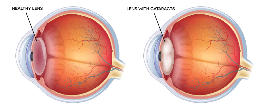
Cataract is an opaque cloudy layer that forms in the lens of the eye. The occurrence starts as proteins in the lens of the eye form clumps, blocking the lens from producing clear images for the retina. It is a condition that gradually develops and obstructs vision. As the cataract becomes bigger, it drives vision to become blurry, hazy, or less colourful and affects normal activities like reading and moving around. Cataract can severely impact quality of life.
Initially, better lighting and prescription eyeglasses helps you in dealing with early cataracts. But, when vision is hampered and obstructs your everyday functioning, you will require to undergo cataract procedure, which is a common, safe, effective method.
Causes
Aging or injuries that distorts the lens tissue in the eyes are the most common causes of Cataracts. When the proteins and fibres in the lens begin to deteriorate, they cause vision to become hazy or cloudy. In other cases, it is possible that some inherited genetic disorders, which are the cause for other health problems, increase the risk of you having cataracts.
They can also be caused by various associated eye conditions, procedure done to the eye in the past and even diabetes. Additionally, it has also been observed that the long-term use of steroid medications too cause cataract.
Symptoms
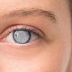
In the beginning the cloudiness caused by a cataract may only affect a portion of the lens. However, when it progresses the lens becomes heavily clouded and distorts the light that comes via the lens.
Clouded vision
Sensitivity to bright light

Poor night vision

Need bright light to read

Halos around lights

Images appear yellow

Double vision in one eye

Diagnosis
Cataract is diagnosed with an all-round and thorough evaluation of the eye by an ophthalmologist. The following tests and examinations are undertaken to accurately diagnose the condition:
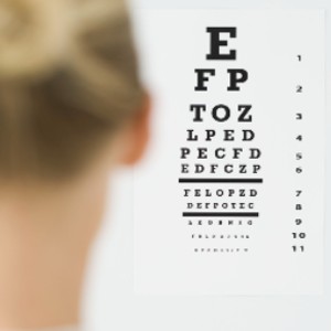
Visual acuity Test
The clarity and sharpness of your vision is assessed with this test. Each eye will individually be examined by making you see letters of different sizes placed at a distance.
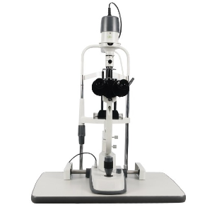
Slit-lamp exam
Different parts of the eye like the cornea, iris, and lens are examined using a special microscope. The lens bending light that enters the eye, during the exam, makes it easier for the ophthalmologist to spot abnormalities.
Read More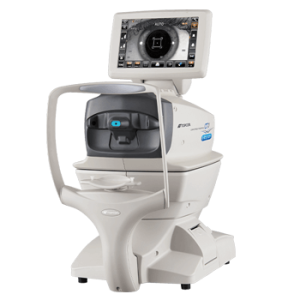
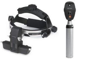
Ophthalmoscopy
Also called fundoscopy, examines the fundus (the inner side of the back in the eye), and evaluates the macula and optic nerve for diseases that could be contributing to the vision loss.
Read MoreTreatments
Cataract has many different procedures that can correct it. At MIOT International Total Eye care, our ophthalmologists ensure to thoroughly diagnose the condition and will explain the best course of correction to be undertaken and why it is optimal.
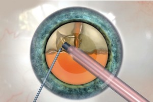
Phacoemulsification
Phacoemulsification is the most common procedure used to remove cataract. It is a simple and swift procedure that is done in not more than an hour. After the lens particles are removed, an intraocular lens is implanted and positioned into the lenses natural capsule. It can be done under minimal sedation, i.e. local anesthesia (injecting anesthesia around the eye) or topical anesthesia (administering numbing drops into the eye).
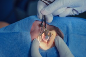
Manual Small Incision Cataract Procedure
An alternative to Phacoemulsification, this procedure uses a small incision that is enlarged through which the damaged eye lens is removed. The capsule of the lens is left behind for the Intra-Ocular Lens (IOL) to be implanted. Many IOL’s are available and serve a various number of purposes. For example some can block out UV light, whereas Multifocal or Trifocal lens provide both near and distant vision correction.
Recent innovations with Intraocular lens (IOL) technology have made micro-incision cataract procedure a very safe and high effective procedure, as only an incision less than 2mm is required to perform the procedure.
Types of Intraocular lens (IOL)
Clearer Vision is Just a Click Away!
Get yourself cared by the most experienced specialists