
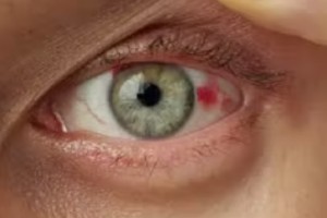
Corneal Diseases
![]()
Cornea is the clear layer in front of the iris and pupil which protects the iris and lens and helps focus light on the retina. Corneal diseases can occur due to infections or damage to the cornea and can lead to visual deterioration and even complete vision loss. It is safer to evaluate the eyes on a regular basis and seek treatment at the first signs.
The Most common type of corneal diseases are:
Diagnosis
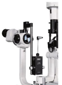
Slit-lamp exam
A slit lamp exam is a routine procedure where a doctor shines a light into the eye to look for injuries or diseases..
Read More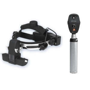
Ophthalmoscopy
It is a simple, painless test to measure the thickness of your cornea -- the clear window at the front of the eye. Corneal thickness has the potential to influence on eye pressure readings.
Read More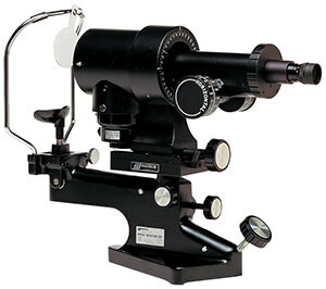
Keratometry
This test is used to measure the anterior corneal curvature of the eye with the help of keratometer. This helps to determine the Differences in power across the cornea which helps to determine any abnormalities in the cornea.
Read MoreTreatments
Treatment is tailored to the individual disease or patient. Treatments might include medications or laser procedure depending on the condition.

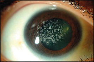
Infections
Treated with medicated eye drops (antibiotics, antivirals, and antiparasitics) and, in some cases, oral medication. Herpetic stromal keratitis is a recurring swelling that develops after a herpes eye infection and is managed with anti-inflammatory steroid eye drops.
Abrasion
Might require temporary patching or a bandage contact lens, depending on the cause and extent of the injury.
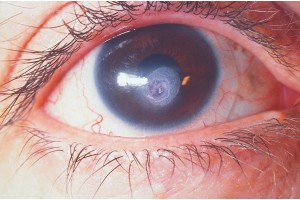
Corneal Perforation
Cyanoacrylate Glue with Bandage Contact Lens (BCL) treatment has become a cornerstone in treating severe corneal thinning and perforations that are less than 2.5 mm in size. High efficacy and ease of application makes it an indispensable tool in the corneal surgeon’s basket. It prevents re-epithelialization into the zone of damaged, naked stroma and the development of the critical setting for collagenase production that leads to stromal melting. Cyanoacrylates also have significant bacteriostatic activity against gram-positive organisms. It is used as a definitive treatment or an interim procedure to delay an otherwise emergent surgical repair.
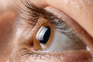
Keratoconus
Cornea can take on a distorted cone shape, which is often managed with special contact lenses. Newer treatments, including corneal crosslinking (riboflavin and ultraviolet-A) and corneal implants, are also options. Advanced keratoconus diseases are treated with specialized contact lenses.
Dystrophies and degenerative corneal disorders
Chronic swelling from Fuchs’ dystrophy or other conditions that damage the cornea’s endothelial cells are managed initially with salty eyedrops or ointments that help prevent accumulation of fluid within the cornea. If the condition worsens, an endothelial lamellar keratoplasty (a type of partial thickness transplant surgery) may be indicated.
Autoimmune disorders
Best treated by addressing the underlying disease. Corneal involvement is often managed with anti-inflammatory eyedrops such as steroids; however, steroid-sparing immune-modulating medications are sometimes preferable, particularly when other parts of the body are also involved.


Pterygium
It is a growth on the cornea’s surface; this is most commonly seen after chronic sun exposure. They can be removed surgically if they become bothersome. Cancers of the surface of the eye are managed with surgery or in some cases, topical chemotherapy eyedrops or injections. The most common procedure performed is Pterygium Excision with conjunctival Autograft.
Clearer Vision is Just a Click Away!
Get yourself cared by the most experienced specialists