
Diabetic Retinopathy
![]()
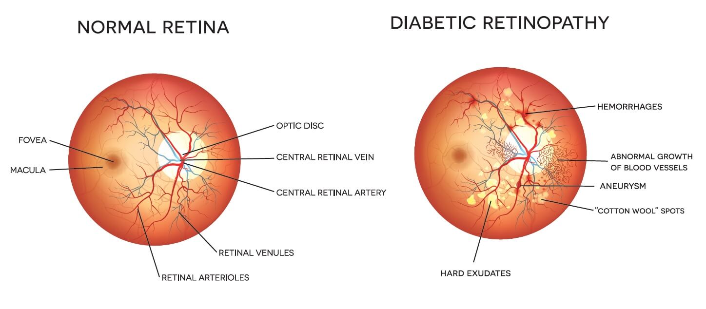
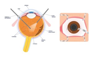
Diabetic retinopathy is a condition that is caused when the small blood vessels in the retina are damaged. Diabetes with irregular control of blood sugar is the main cause. It occurs as high blood sugar levels damage the blood vessels. In this condition the blood vessels can swell, leak and can even close, stopping flow of blood through the eye. In some cases, new blood vessels can even grow on the retina.
Initially, diabetic retinopathy may reveal no symptoms or appear only as mild visionary ailments. But without proper check-ups and intervention, when undiagnosed, it will lead to blindness. The condition can affect both type 1 and type 2 diabetics. The likelihood of Diabetic Retinopathy is higher in uncontrolled blood sugar. Although, mild cases can be treated with careful management of diabetes, advanced cases may have to be treated using laser treatment or procedure.
Causes
Diabetic retinopathy is directly the effect of uncontrolled high blood sugar levels because of diabetes. Over a course of time, excessive sugar levels in your blood damage your retinal blood vessels – This causes the retina to be deprived of blood and nutrients.
Everyone is familiar with the cause and effect of diabetes and how it can damage blood vessels in the body. As this is happening, it affects the eyes when the blood vessels of the eyes are damaged, and as a result they leak fluid or bleed. As a reactionary measure to make up for blocked blood vessels, the eyes generate new blood vessels, but don’t work well and leak or bleed easily.
Symptoms
Diabetic Retinopathy almost has no symptoms in the early stages. It is better to check one’s vision if they have diabetes to intervene before it gets serious. But when people do have symptoms, they can notice:
Floating Spots

Dark Strings floating

Blurred Vision

Fluctuating Vision

Dark areas in vision

Vision loss

Colour Blindness

Difficulty colors

Diagnosis
Diabetic Retinopathy is a direct complication caused by the presence of diabetes. It affects up to 80 percent of those who have had diabetes for 5 years or more. The longer you are diabetic, the greater are your chances of being affected. Since it is a gradual condition, regular eye check-ups are required to identify its development and proceed with the right course of treatment. The following tests and examinations are undertaken to accurately diagnose the condition:
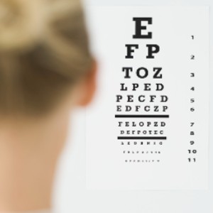
Visual acuity Test
The clarity and sharpness of your vision is assessed with this test. Each eye will individually be examined by making you see letters of different sizes placed at a distance.

Dilated eye exam
Eye drops are placed in the eyes to widen (dilate) your pupils to allow the specialist to get a better view inside your eyes. Any abnormalities in the inside and outside parts of your eyes are carefully examined..

Optical coherence tomography (OCT)
OCT provides cross-sectional images of the retina that show the thickness of the retina. This will help determine how much fluid, if any, has leaked into retinal tissue.
Read More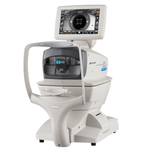
Optical coherence tomography (OCT)
It enables the ophthalmologist to take a closer look at the retina. During the test, the machine scans the retina and provides detailed images of its thickness. Helping the doctor look for and measure the swelling of the macula.
Read MoreTreatments
Treatment for diabetic retinopathy can vary based on the extent of the disease. But regardless of the presence of the disease controlling diabetes with prescribed medication, regular exercise, diet and healthy habits can reduce the risk of it.
At MIOT International Total Eye care, our ophthalmologists ensure to thoroughly diagnose the condition and will explain the best course of correction to be undertaken and why it is optimal.

Medical management
Injection of anti-VEGF medication into the eye can help to reduce swelling of the macula, slowing vision loss and even improving vision, it also helps in reducing the new vessel formation. Steroid injections are also an alternate option used to reduce macular swelling.
Laser procedure
As and when diabetic retinopathy advances, blood vessels can leak blood and fluid into the retina, causing macular edema. Laser treatment helps in stopping this leakage. Focal laser photocoagulation uses a laser to target a specific leaky vessel in the macula to keep macular edema / swelling in check.
When the blood vessel growth in the retina is widespread it is treated by creating a pattern of scattered laser burns across the retina. After which, abnormal blood vessels either shrink or disappear.
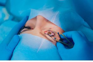
Vitrectomy
Vitrectomy is a procedure that removes scar tissue and blood from the vitreous fluid of the eye. In this procedure a tiny incision in made in the eye to remove blood from the middle of the eye (vitreous humor) as well as scar tissue that’s tugging on the retina. It is a simple and safe procedure that can be performed with local or general anaesthesia.
Treatment can only slow or stop the progression of diabetic retinopathy. It is not the final cure as diabetes is a lifelong condition and future retinal damage and vision loss are still possible. Regular check-up and eye exams combined with additional treatment will be required to keep it under control and to prevent further damage.

Clearer Vision is Just a Click Away!
Get yourself cared by the most experienced specialists